


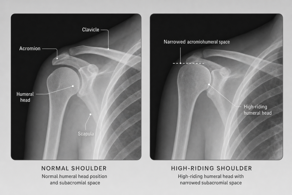
High-Riding Humeral Head on X-Ray: What It Can Mean
A high-riding humeral head on x-ray is a radiographic finding rather than a stand-alone diagnosis. It means the ball of the upper arm bone appears to sit higher than expected relative to the acromion on shoulder imaging. On its own, that does not confirm one specific condition, but it can point toward rotator cuff dysfunction, chronic cuff tearing, or cuff tear arthropathy and should be interpreted together with symptoms, examination findings, and imaging context.
When a shoulder x-ray report mentions “superior migration of the humeral head” or a narrowed “acromiohumeral interval,” many patients are left wondering whether the finding automatically means a torn rotator cuff or surgery. Usually, it does not. It is better understood as an imaging clue that helps narrow the diagnosis. For a broader overview of shoulder pain causes and evaluation, readers can start with our general shoulder pain hub.
What a High-Riding Humeral Head Means on an X-Ray
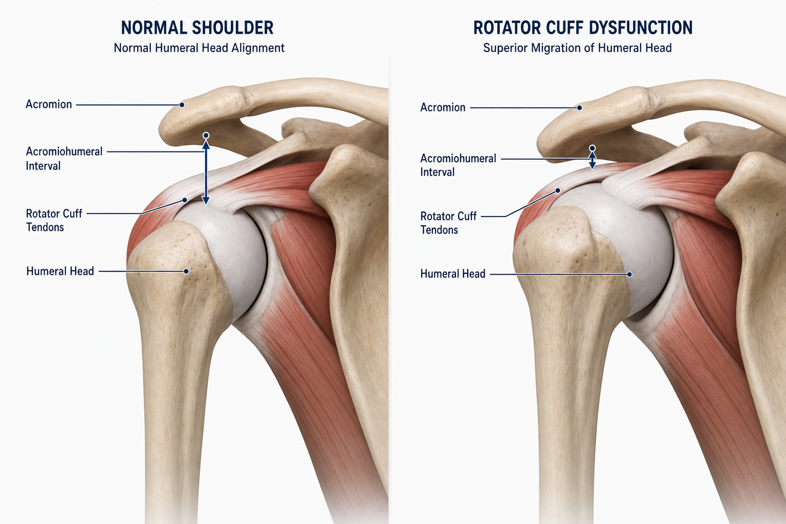
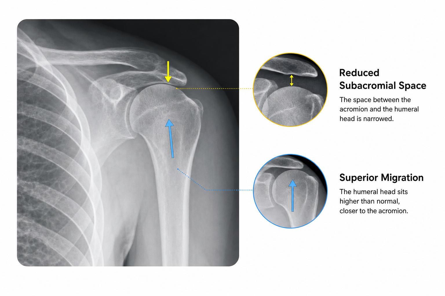
On a standard shoulder x-ray, clinicians look at how the humeral head sits relative to the socket and the acromion above it. The space between the inferior acromion and the top of the humeral head is called the acromiohumeral interval. A reduced interval can reflect superior humeral head migration, especially in chronic rotator cuff disease, although x-ray positioning can influence the measurement.
That is why “high-riding humeral head meaning” is best answered this way: it is an imaging sign that may reflect abnormal shoulder mechanics, commonly from rotator cuff dysfunction, but it is not the final diagnosis by itself. A 2025 systematic review found that reduced acromiohumeral distance tends to be associated with greater rotator cuff tear severity, especially multi-tendon and massive tears, while also noting heterogeneity across studies. Systematic review on acromiohumeral distance and rotator cuff tears

Why the Humeral Head Can Shift Upward
The rotator cuff helps keep the humeral head centered in the socket during motion. When the cuff is substantially torn or chronically weakened, the balance of forces around the shoulder changes, and the deltoid can pull the humeral head upward more than normal. Reviews of shoulder biomechanics and imaging describe this loss of force balance as a major explanation for superior migration in chronic cuff disease. Narrative review on superior migration of the humeral head
Still, rotator cuff tearing is not the only possible reason. Chronic cuff tear arthropathy, prior surgery, capsular contracture, some arthritic patterns, and other changes in shoulder mechanics can also affect apparent humeral head position. That is one reason patients often need more than just the x-ray report to understand what the finding means.
For readers comparing tear patterns, our page on partial vs. full-thickness rotator cuff tears explains how tear size and tendon involvement can change treatment planning.
Common Conditions Linked to a High-Riding Humeral Head
Conditions commonly associated with a high-riding humeral head on x-ray include:
- Large or massive rotator cuff tears: These are among the most common associations, particularly when more than one tendon is involved.
- Chronic rotator cuff disease: Long-standing cuff dysfunction can gradually allow superior migration of the humeral head.
- Rotator cuff arthropathy: This is a specific combination of chronic cuff failure and arthritic joint change.
- Advanced shoulder arthritis with altered mechanics: In some patients, arthritis contributes to abnormal positioning, especially when cuff tissue is also compromised.
- Pseudoparalysis: Severe cuff dysfunction can be associated with marked loss of active elevation, although pseudoparalysis is a clinical finding rather than an x-ray diagnosis.
If your question is whether a high-riding humeral head rotator cuff tear connection is real, the answer is yes: it is a recognized association, particularly with chronic larger tears, but it is not an absolute rule in every patient.
Symptoms That Matter Along With the X-Ray Finding
The same x-ray finding can matter very differently depending on symptoms and function. Important accompanying symptoms may include:
- Shoulder pain with reaching or overhead activity
- Weakness lifting the arm
- Difficulty raising the arm actively
- Night pain
- Grinding, catching, or mechanical symptoms
- Loss of function after an injury or gradual decline over time
Patients with marked weakness, loss of active elevation, or worsening function generally warrant closer evaluation than those with a mild incidental x-ray finding and minimal symptoms.
What Imaging or Tests May Be Needed Next
X-rays show bone alignment well, but they do not directly show the rotator cuff tendons. Depending on the history and exam, an MRI may be ordered to evaluate tendon integrity, muscle quality, retraction, arthritis, and whether the pattern looks repairable. MRI is often useful, but it is not automatically necessary for every patient if the diagnosis and treatment path are already clear clinically.
If a report feels vague or hard to interpret, a shoulder MRI second opinion can help connect the images to realistic treatment options and expectations.
When Non-Surgical Treatment May Help
Non-surgical treatment can help many patients, especially when pain is manageable, function is partly preserved, or goals do not require immediate surgery. AAOS guidance on rotator cuff injuries emphasizes that treatment decisions depend on factors such as age, activity level, tear cause, tear size, health status, pain, and functional impact rather than a one-size-fits-all rule. AAOS plain-language summary on management of rotator cuff injuries
Common non-surgical approaches may include:
- Physical therapy: A program focused on motion, deltoid function, scapular control, and remaining cuff strength can improve pain and use of the arm in selected patients.
- Activity modification: Reducing aggravating overhead or repetitive loading can calm symptoms.
- Medications or injections: These may sometimes be used for symptom relief, depending on the diagnosis and patient factors.
- Observation with follow-up: In some lower-demand or minimally symptomatic patients, careful monitoring may be reasonable.
For conservative care options, see our pages on non-surgical shoulder treatment and shoulder physical therapy.
PRP is sometimes discussed for shoulder pain, but current AAOS clinical practice guidance does not support routine use of platelet-rich plasma for rotator cuff tendinopathy or partial tears. That means any discussion of PRP should be individualized and should not be presented as proven standard treatment for this x-ray finding. AAOS clinical practice guideline on rotator cuff injuries

When Surgery Becomes Part of the Discussion
Surgery may enter the discussion when pain remains limiting despite appropriate non-surgical care, when weakness or function loss is substantial, or when imaging shows pathology that is unlikely to do well without operative treatment. The procedure depends on the cause of the superior migration rather than the x-ray phrase alone.
- Rotator cuff repair or arthroscopic treatment: Some patients with repairable tears may be candidates for surgery aimed at restoring the cuff and shoulder mechanics.
- Reverse shoulder replacement: In patients with cuff tear arthropathy or irreparable cuff failure with major dysfunction, reverse shoulder arthroplasty may be considered.
- Other individualized procedures: Prior surgery, arthritis severity, and tissue quality can change the recommendation.
To learn more about procedure pathways, readers can review reverse shoulder replacement for cuff arthropathy if cuff tear arthropathy is suspected.
Questions to Ask at Your Shoulder Evaluation
Helpful questions include:
- What is the most likely cause of the superior humeral head migration in my case?
- Do my symptoms fit a rotator cuff tear, cuff tear arthropathy, arthritis, or something else?
- Would MRI change treatment planning for me?
- Is the rotator cuff likely repairable if torn?
- What non-surgical treatment is reasonable first?
- What would make surgery more likely to help?
Our first shoulder specialist visit checklist can help you gather questions and prior imaging before the visit.
A Hypothetical Example: Putting the Finding in Context
Common Mistakes Patients Make After This Finding
- Assuming the x-ray phrase is the full diagnosis: It is not.
- Assuming surgery is automatic: Many patients are first managed without surgery, depending on symptoms and goals.
- Ignoring major weakness or function loss: These symptoms can matter as much as the image.
- Relying only on internet definitions: Imaging language needs clinical context.
- Expecting any one treatment to fit everyone: Treatment planning is individualized.
Frequently Asked Questions
What does a high-riding humeral head on an X-ray mean?
It means the humeral head appears to sit higher than expected on the x-ray, reducing the space between it and the acromion. It is a radiographic sign that can suggest abnormal shoulder mechanics, commonly related to rotator cuff dysfunction.
Does a high-riding humeral head mean I have a rotator cuff tear?
Not always, but rotator cuff tearing is one of the most common associations, especially when the tear is chronic or large. The finding still needs to be interpreted with symptoms, examination, and sometimes MRI.
Can shoulder arthritis cause a high-riding humeral head?
Yes, especially when arthritis is combined with rotator cuff deficiency, as in cuff tear arthropathy. Arthritis alone and other mechanical changes can also affect how the humeral head appears on imaging.
Do I need an MRI after my X-ray report mentions superior migration?
Sometimes, but not always. MRI is often helpful when a clinician needs to assess the rotator cuff tendons, muscle quality, and joint surfaces in more detail, but the decision depends on the clinical picture.
Can a high-riding humeral head improve with physical therapy?
Physical therapy may improve pain and function in some patients, even if the underlying x-ray appearance does not fully normalize. Whether therapy is enough depends on the cause, severity, and functional goals.
When does a high-riding humeral head lead to reverse shoulder replacement?
That discussion usually comes up when there is irreparable rotator cuff dysfunction with major loss of function, often along with cuff tear arthropathy or arthritic joint change. The x-ray wording alone is not enough to make that decision.
Should I see a shoulder specialist if urgent care found this on an X-ray?
If you also have ongoing pain, weakness, limited motion, or uncertainty about the report, specialist follow-up is reasonable so the finding can be interpreted in the right clinical context.
Update Log
- 2026-10-16: Article generated for publication.