

Subchondral Cysts on Shoulder MRI Explained
If your shoulder MRI report mentions subchondral cysts or subchondral cystic change, it is natural to have questions about what that finding means. This guide explains what subchondral cysts are, where they appear, common causes, and how this MRI finding is interpreted together with your symptoms, physical exam, and other imaging.
What Subchondral Cysts on a Shoulder MRI Actually Mean
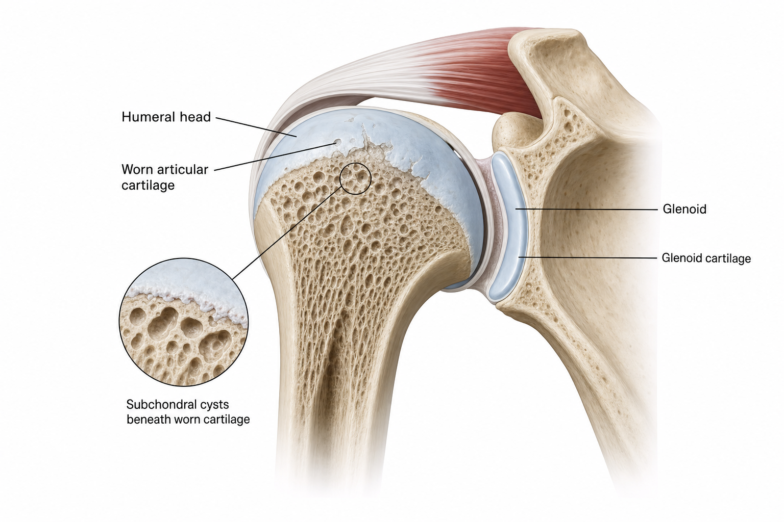
The term subchondral means beneath the cartilage. Subchondral cysts are small fluid-like spaces that develop in bone just below the joint surface. On MRI, they appear as rounded areas of signal change in places such as the humeral head or glenoid.
These cysts are not the same as paralabral cysts, ganglion cysts, or the more superficial cysts that may occur around the AC joint. In general, they are interpreted as part of broader structural change in the shoulder joint rather than as an isolated disease. That is why understanding the causes of shoulder pain starts with reading the MRI in context, not just reacting to a single phrase in the report.
Where These Cysts Appear in the Shoulder Joint
In the glenohumeral joint, subchondral cysts are often described in the humeral head or the glenoid. When arthritis or cartilage wear is more diffuse, they may be present on either side of the joint. When the MRI describes humeral head cystic change, the exact location matters because some cyst patterns are more associated with age-related change, while others can be associated with rotator cuff abnormalities.
Location also helps distinguish this finding from other cyst types. For example, AC joint cysts and paralabral cysts may appear near the shoulder but have different causes and implications. If your report is difficult to follow, a shoulder MRI second opinion can help translate the wording into plain language.
Common Causes: Arthritis, Cartilage Wear, and Cuff Disease
Common reasons a shoulder MRI may mention subchondral cystic change include:
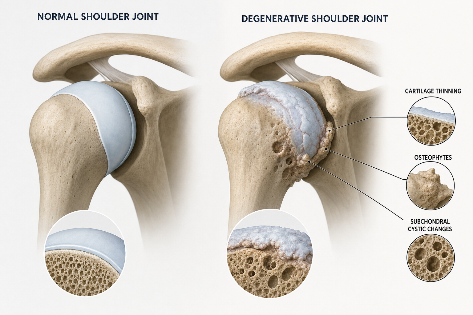
- Glenohumeral osteoarthritis: As cartilage thins and the joint surface changes, the bone beneath the cartilage can also change. Subchondral cysts are one of several degenerative findings that may appear alongside osteophytes, cartilage loss, and joint-space loss.
- Focal cartilage wear or osteochondral injury: Localized cartilage damage can be associated with more focal subchondral bone change.
- Rotator cuff-related degeneration: Some humeral head cyst patterns are linked to rotator cuff disease, especially near tendon insertion sites, while other patterns are more related to aging and not necessarily a tear. This is why cuff-related MRI findings should be interpreted carefully rather than assumed to mean automatic surgery. Readers comparing tear patterns may also find partial versus full-thickness rotator cuff tears helpful.
- Prior instability or earlier joint injury: Over time, altered loading after instability or injury can contribute to focal cartilage and subchondral change.
If arthritis is suspected, plain X-rays remain important because they help show joint-space narrowing, bone spurs, and overall joint architecture in a way MRI wording alone cannot. For arthritis-focused evaluation, see shoulder arthritis care.
Do Subchondral Cysts Always Cause Shoulder Pain?
Not always. Some subchondral cysts are incidental imaging findings, especially when they are small and the shoulder is otherwise functioning fairly well. In those situations, the cyst itself may not be the pain generator.
Symptoms are more likely to come from the condition associated with the cystic change, such as arthritis, stiffness, cartilage wear, or rotator cuff pathology. Larger or more numerous cysts, especially when seen alongside cartilage thinning or other degenerative changes, may suggest a more advanced joint process, but they still do not determine treatment on their own.
How a Shoulder Specialist Interprets This MRI Finding
A shoulder MRI is only one part of the evaluation. A specialist usually asks: How much cartilage is left? Is the rotator cuff intact? Is there stiffness or weakness on exam? Do X-rays show joint-space loss or a pattern suggesting cuff tear arthropathy? Is there a high-riding humeral head on X-ray that changes the differential?
This approach is important because the same phrase in a radiology report can mean different things in different patients. For one person, it may reflect mild age-related degeneration. For another, it may fit glenohumeral osteoarthritis or cuff-related arthropathy that changes treatment planning. If you have ongoing pain and an unclear report, a shoulder-focused review can help determine whether you mainly need reassurance, non-surgical care, or a discussion of more advanced options.

When Treatment Is Non-Surgical and When Advanced Care Is Needed
Treatment is aimed at the underlying shoulder condition, not at removing the cyst itself. When symptoms are mild or moderate, non-surgical treatment is often the first step. Options may include activity modification, anti-inflammatory measures when appropriate, and a structured rehabilitation plan through non-surgical shoulder treatment.
Depending on the diagnosis, physical therapy may help improve motion, shoulder mechanics, and function. Injections may also be considered in selected cases, particularly for symptomatic arthritis. If PRP is discussed, it should be presented cautiously: PRP is not a direct treatment for a subchondral cyst, and evidence for shoulder conditions is mixed and condition-specific rather than uniformly established.
When pain, loss of motion, structural degeneration, or cuff-related arthropathy are more advanced, surgical options may enter the discussion. Those options depend on the diagnosis and may range from arthroscopic procedures in selected cases to shoulder replacement when joint disease is advanced. The goal is to match treatment to symptoms, function, imaging, and patient goals rather than to the MRI phrase alone.
Red Flags That Mean You Should Book a Shoulder Evaluation
Consider a formal shoulder evaluation if your MRI mentions subchondral cystic change and you also have:
- Persistent or worsening shoulder pain
- Marked stiffness or progressive loss of motion
- Weakness lifting the arm or reaching overhead
- Night pain that regularly interrupts sleep
- A history of prior dislocation, major injury, or prior shoulder surgery
- An MRI report that also mentions cartilage loss, cuff tearing, or advanced degenerative changes
If symptoms began after major trauma, or if you have sudden severe weakness, fever, visible deformity, or inability to use the arm, urgent evaluation is more appropriate than routine online education.
Questions to Ask After an MRI Report Mentions Cystic Change
Helpful questions for your visit include:
- Is this cystic change most consistent with arthritis, focal cartilage wear, or rotator cuff-related degeneration?
- Do my X-rays show joint-space loss or cuff tear arthropathy?
- Are the cysts likely incidental, or do they fit the main cause of my pain?
- What non-surgical treatments make sense for my diagnosis?
- What findings would make surgery worth discussing?
If you are preparing for a specialty visit, the first shoulder specialist visit checklist may help you organize questions, prior imaging, and symptom history.
Frequently Asked Questions
What does subchondral cystic change in the shoulder mean on MRI?
It means there are small fluid-like spaces in bone beneath the joint cartilage. On shoulder MRI, this usually suggests an underlying structural process such as cartilage wear, osteoarthritis, or in some cases cuff-related degeneration, rather than a separate diagnosis by itself.
Does a shoulder subchondral cyst mean I have arthritis?
Not always, but arthritis is a common explanation. Some cyst patterns can also be related to localized cartilage damage, aging-related bone change, or rotator cuff pathology. The location and the rest of the imaging matter.
Can subchondral cysts be caused by a rotator cuff tear?
Some humeral head cysts are associated with rotator cuff disease, especially near tendon attachment sites. However, not every humeral head cyst means there is a cuff tear, and some are seen with aging even when the cuff is intact.
Do subchondral cysts in the humeral head go away?
They typically are not the direct target of treatment. The focus is on managing the underlying condition causing pain or joint stress, such as arthritis, stiffness, or rotator cuff-related problems.
Is surgery needed for subchondral cysts in the shoulder?
No. Surgery is not based on the cyst alone. It depends on your symptoms, exam findings, response to non-surgical care, rotator cuff status, and the degree of overall joint degeneration.
When should I see a shoulder specialist after an MRI report?
You should consider an evaluation if you have ongoing pain, stiffness, weakness, night pain, loss of function, or a report that mentions cystic change along with arthritis or rotator cuff abnormalities.
Sources
Update Log
- 2026-10-21: Article generated for publication.